Today's hours: Closed

 네이버지도
 카카오맵
 구글지도
 TMAP

Cancer Screening
Gynecological, Breast, and Thyroid Comprehensive Screening
We conduct step-by-step examinations of the cervix, ovaries, endometrium, breasts, and thyroid gland using ultrasound and cytology.
Ultrasound
Ultrasound Exams: Be Sure to Get One in These Situations!
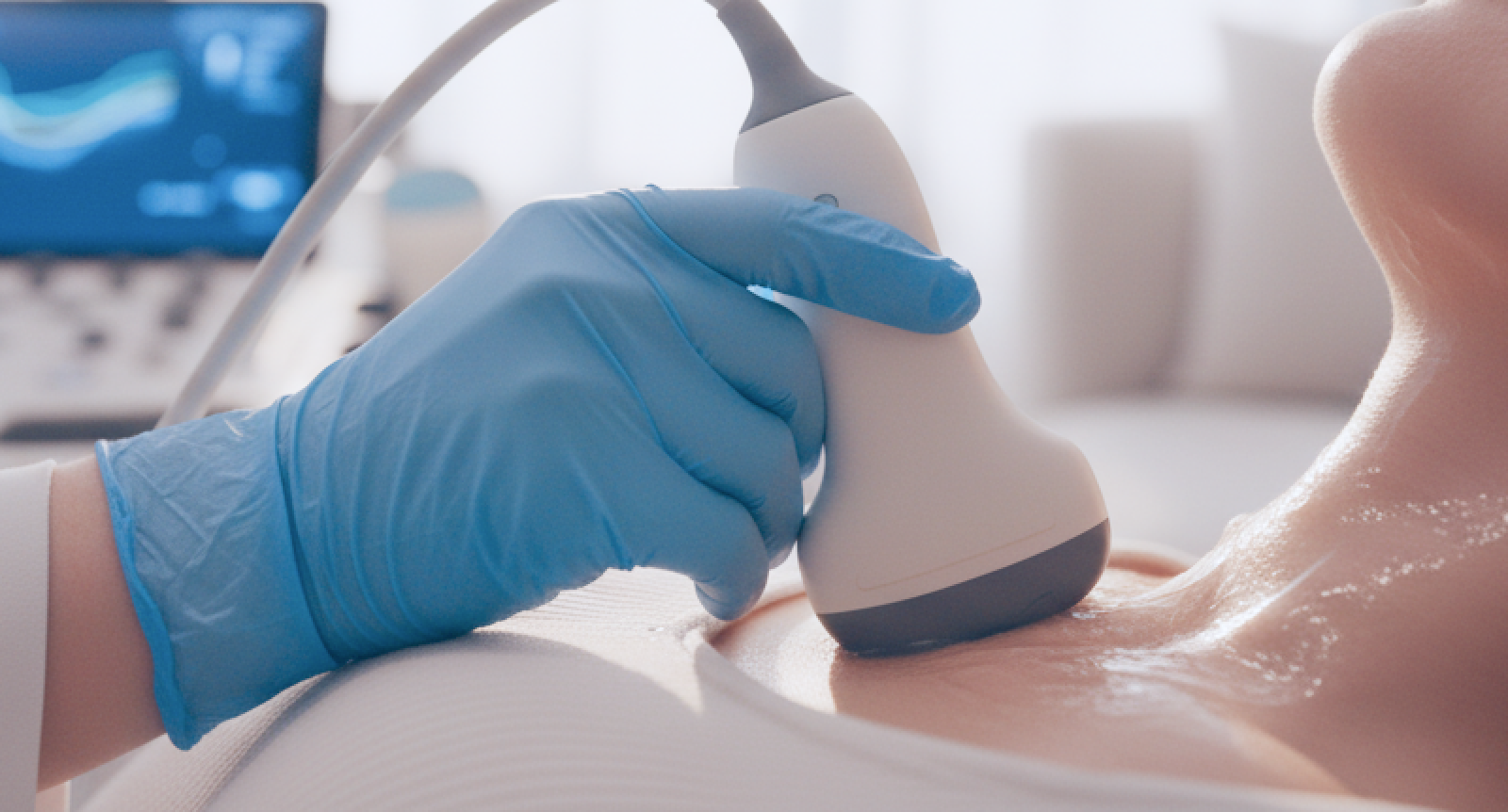
Ultrasound examinations, which involve no radiation exposure and have a wide range of applications, are widely used for the early diagnosis of various diseases, including cancer. Ultrasound examinations are said to be safe and have a very broad diagnostic range, covering organs, muscles, and blood vessels. They are gaining recognition as the most fundamental and reliable method of diagnostic imaging, and another appealing feature of ultrasound is its ability to provide real-time feedback.
Since the images are displayed in real time, patients can hear an explanation of the results immediately and receive a prompt diagnosis.
Environmental factors can significantly increase the risk of sudden death due to heart disease, such as myocardial infarction; the heart and blood vessels are the most critical areas to monitor within the normal range. Sudden vasoconstriction puts strain on the heart, leading to fatal conditions such as acute myocardial infarction. Since heart disease often presents without specific symptoms, it is advisable for people in their 40s and older to have their heart health checked regularly. An ultrasound is a crucial test that allows for the assessment of the heart’s sustained contraction and relaxation, as well as the integrity of the heart walls and valves.
Thyroid Ultrasound
- If you have a hoarse voice or persistent vocal cord paralysis
- If you have a history of radiation therapy to the head and neck region
- If you have a family history of thyroid cancer
- If a thyroid nodule (lump) has suddenly grown larger
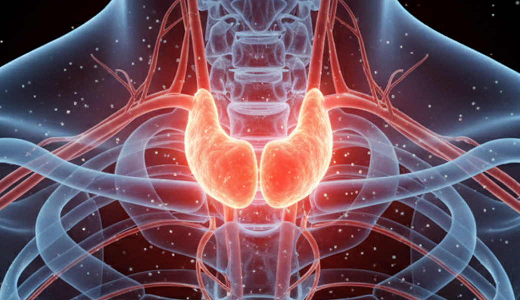
Abdominal Ultrasound
- Abdominal pain or persistent indigestion
- When blood tests show elevated liver enzyme levels or hemoglobin levels
- For women, when abdominal pain or sudden changes in the lower abdomen raise suspicion of uterine fibroids
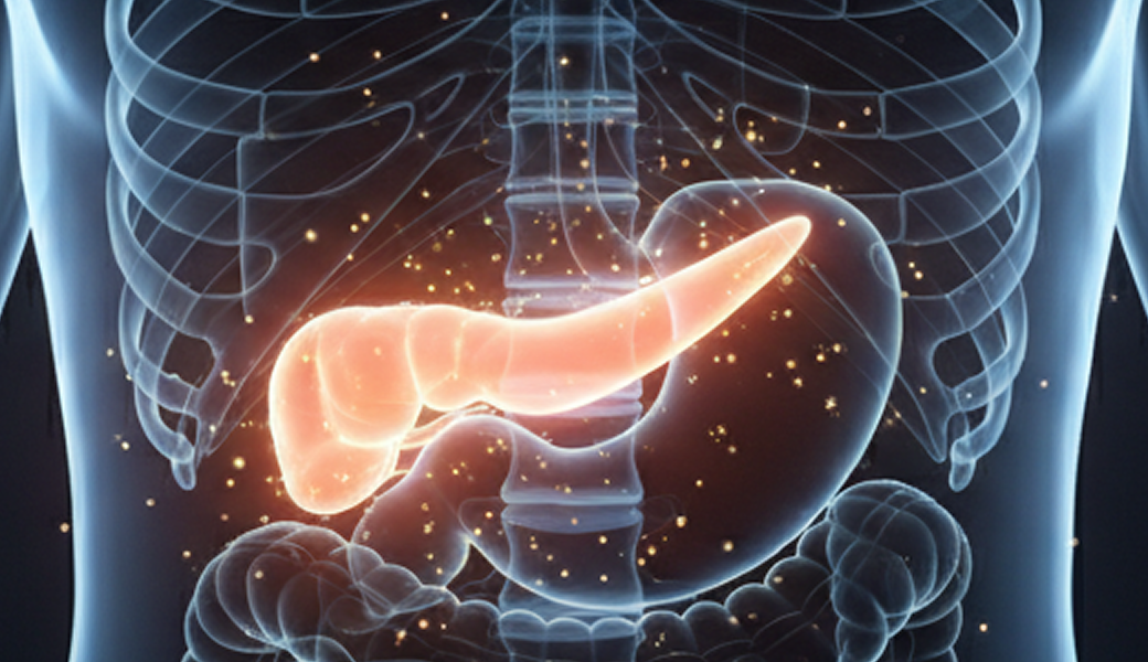
Breast Ultrasound
- If you are concerned about breast cancer or feel a lump
- If you experience nipple discharge or a change in skin color
- When examining implants or breast tissue following breast augmentation surgery
- If you have dense breasts, even if no abnormalities are found, this test may be used for additional screening

For women, ultrasound screening is not an option but a necessity. To improve the accuracy of early breast cancer detection, mammography—which uses X-rays—and breast ultrasound should be performed together. Most women in Korea have dense breasts with tightly packed glandular tissue. Dense breasts make it difficult to visualize the tissue on a mammogram, so standard screening methods are not well-suited for them. This phenomenon is particularly pronounced in young women in their 30s with dense breasts.
Gynecological Ultrasound
- Uterine fibroids, adenomyosis, endometriosis, ovarian disorders, and pelvic inflammatory diseases can be detected through regular gynecological ultrasound exams.
- Uterine fibroids, whose incidence is rising among young women, can also be diagnosed through a thorough ultrasound exam, and if detected early, they can be treated without surgery.
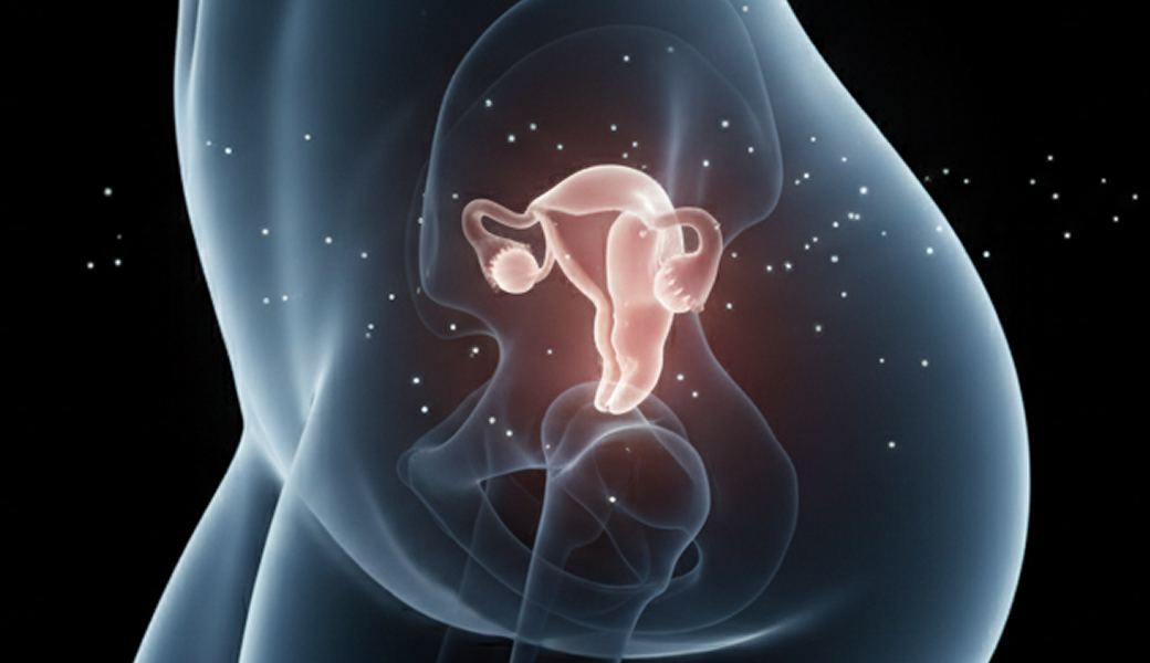
Cervical Cancer
Cervical Cancer
The cervix is the part of the body that connects the vagina to the uterus; it plays a crucial role in childbirth, and this is a type of female genital cancer that occurs in the cervix.
Causes of Cervical Cancer
Human Papillomavirus (HPV) Infection Is the Primary Cause
HPV is a virus that very commonly infects the skin around the anus and genitals in men. Although it is often asymptomatic, once the virus causes a condition, it does not go away easily without treatment.
Summary of Information on HPV (Human Papillomavirus)
| Women's Experience with Sexually Transmitted Infections | Eight out of every 10 sexually active women become infected at least once in their lifetime without even realizing it. |
| HPV Type Diversity | Currently, approximately 130 different types of HPV have been identified. |
| High-Risk Infection Outcomes (Related to Lifestyle Habits) | Recurrent infections with high-risk viruses in some women can lead to various genital diseases, such as cervical cancer, genital warts (condyloma or condyloma acuminatum), vulvar cancer, and vaginal cancer. |
| Characteristics of the Infection | Since most people do not experience any noticeable signs or symptoms, they become infected without realizing it and spread the infection to others. |
| Diseases by Major Type | Approximately 70% of cervical cancer cases are caused by HPV types 16 and 18.- More than 90% of genital wart cases are caused by HPV types 6 and 11. |
Cervical Cancer Screening

Cervical Pap Smear
- The most widely used screening method for the early diagnosis of cervical cancer, which helps reduce its incidence
- Women who are sexually active should undergo annual screenings

Human Papillomavirus (HPV) Test
- To compensate for the high risk associated with cervical specimens, a molecular genetic test is performed as an adjunct to confirm the presence or absence of HPV infection, and patients are classified into high-risk and low-risk groups.
- High-risk types: 16, 18, 26, 31, 33, 35, 39, 45, 51, 52, 53, 56, 58, 59, 66, 68, 69, 70, 73, 82
- Low-risk group: 6, 11, 40, 42, 43, 44, 54, 61 → The HPV types that primarily cause cervical cancer are 16 and 18

Cervical Dilatation and Dilation
- A test designed to compensate for the low sensitivity of cervical cancer screening and reduce the false-positive rate of Pap smears
- A diagnostic method in which a specialist identifies abnormal areas in images magnified 15 to 20 times during cervical magnification imaging

Tissue biopsy
- If changes are observed during a preliminary examination, they are confirmed through a biopsy.
- A very small sample of cervical tissue is removed and examined under a microscope to make a histological diagnosis.
Treatment for Cervical Cancer
Since regular cervical screenings can detect cervical intraepithelial neoplasia at an early stage, when treatment is most likely to be successful, early detection is of the utmost importance.

Hyperthermia Therapy
Loop electrosurgical excision procedure
- Method for Removing the Entire Lesion Using a Wire Loop
- Diagnosis and treatment are performed in a single procedure using the excised tissue, and the procedure can be performed on an outpatient basis.

Cone Biopsy
Conization
- Resection using a cold knife, carbon dioxide (CO₂) laser, or electrosurgical instrument
- Can be performed for diagnostic and therapeutic purposes

Hysterectomy
- Among the treatment options for cervical intraepithelial neoplasia and cervical cancer, this is the one with the lowest recurrence rate.

Radiation Therapy and Chemotherapy
Cervical Cancer Vaccination
Cervical cancer is the only preventable form of cancer; vaccination to prevent HPV infection is particularly recommended.

HPV Types (16, 18, 6, 11) A quadrivalent vaccine that protects against these four types

9-valent vaccine: 4 HPV types (16, 18, 6, 11) + 5 additional types (31, 33, 45, 52, 58)
National Free Vaccination Program
For female adolescents aged 12 to 13
- Two free vaccine doses administered on a 0-month and 6-month schedule
- Quadrivalent vaccine (16, 18, 6, 11)
Ovarian Cancer
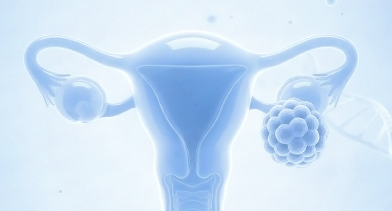
Ovarian Cancer
- This refers to cancer that originates in the ovaries—where eggs, which are germ cells, mature and where female hormones are secreted periodically—and is a type of cancer in women whose incidence has been rising recently.
- Most ovarian cancers progress rapidly and are often diagnosed at an advanced stage, so early detection through regular gynecological exams is crucial.
Ovarian Cancer Diagnosis

Initial Examination
After undergoing tests such as an ultrasound, CA-125, or ROMA blood test, ovarian cancer is suspected

Second Examination
Determining whether a tumor is benign or malignant through tests such as computed tomography (CT) and magnetic resonance imaging (MRI)

Final Diagnosis
Diagnosis is made through histopathological examination following the removal of an ovarian mass via open surgery or laparoscopic surgery.

Endometrial Cancer
Endometrial Cancer
- A condition in which cancer composed of abnormal cells develops in the endometrium
- It is also called “endometrial cancer” because it originates in the endometrium and grows into the muscular layer, which makes up most of the uterus
- Although its incidence is higher in the West than in the East, it has been gradually increasing in Korea as our lifestyle becomes more Westernized
Diagnosis and Treatment of Endometrial Cancer
Symptoms such as vaginal bleeding, postmenopausal uterine bleeding, lower abdominal pain, increased vaginal discharge, and heavy menstrual bleeding may occur

Biopsy
Remove the lesion through procedures such as endometrial curettage and perform a biopsy

Treatment
Most cases are detected relatively early, and radiation therapy is used in conjunction with surgery to prevent recurrence, achieve a cure, and treat metastases.
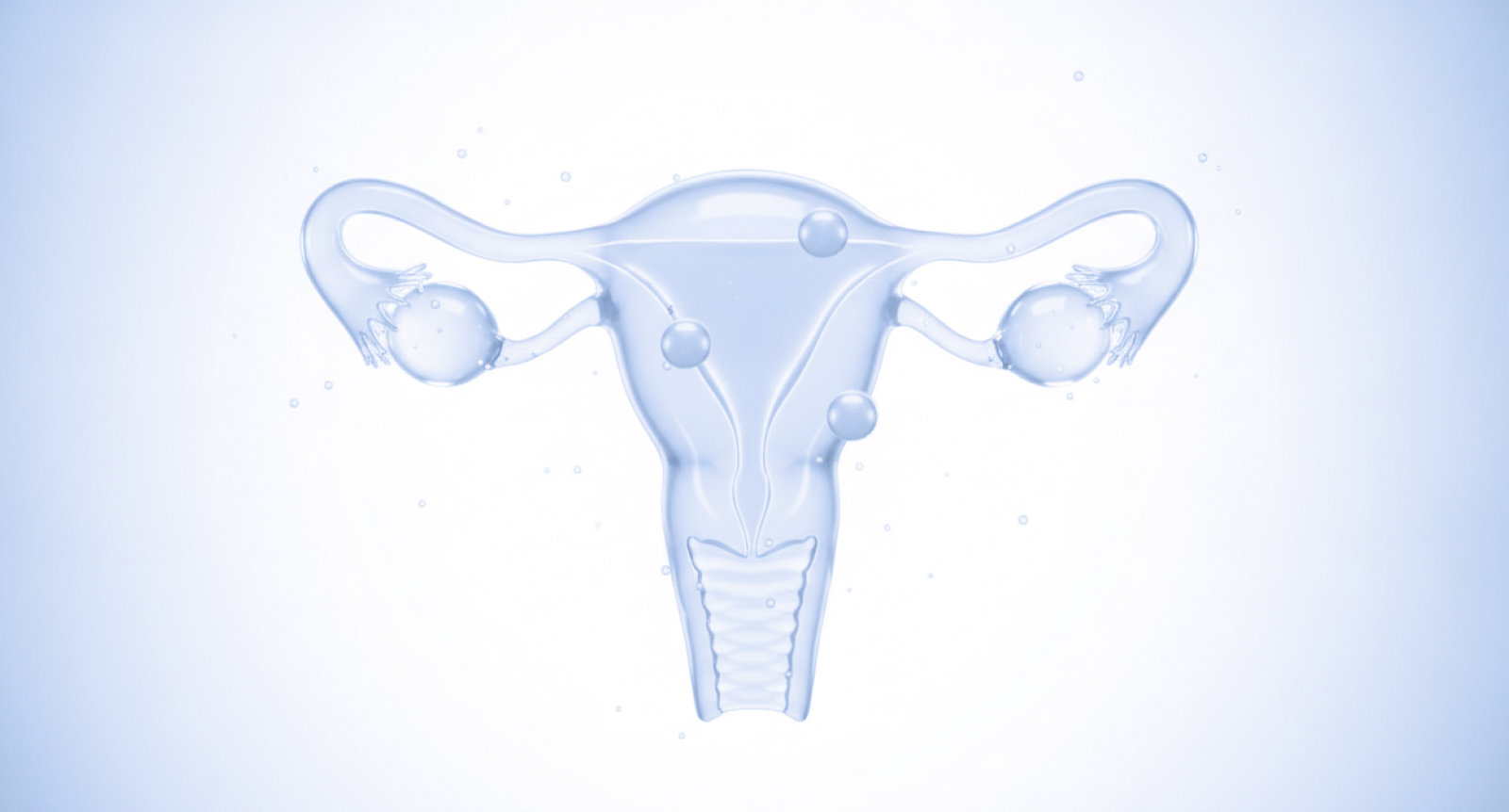
Uterine Fibroids
Uterine Fibroids
Uterine fibroids are benign tumors that develop in the muscular layer of the uterus and are the most common pelvic condition affecting women.
- Can occur at any age
- Vary in size and number
- Associated with abnormal uterine bleeding (such as heavy menstrual flow and irregular bleeding), pelvic pain, frequent urination due to pelvic pressure, constipation, infertility, and recurrent miscarriages
Diagnosis of Uterine Fibroids

Pelvic exams and ultrasounds are the most common

Hysteroscopy

To determine the location of the site prior to surgery, magnetic resonance imaging (MRI) or computed tomography (CT) is used
Treatment for Uterine Fibroids

Follow-up
If symptoms are absent or mild, monitor the condition with regular ultrasound examinations

Drug Therapy
Methods for reducing the size of fibroids using estrogen blockers and other treatments

Surgery
Surgery for severe symptoms

Moderation
Depending on whether the patient wishes to become pregnant, as well as the size and shape of the fibroids and the patient’s age, either the fibroids alone or the entire uterus may be removed.

Breast Cancer
Breast Cancer Screening
The Most Common Cancer Among Women
Breast cancer currently occurs most frequently in women in their 40s and 50s, and the incidence among women in their 20s and 30s is also gradually increasing. Fortunately, however, if breast cancer is detected early, a cure rate of over 95% can be expected.
Breast Cancer Diagnosis
Like other cancers, breast cancer is difficult to detect in its early stages because it does not present any characteristic symptoms; therefore, regular checkups and screenings, as well as breast self-exams, are important. If you notice any symptoms or abnormalities, it is advisable to see a specialist immediately and undergo thorough testing.

Breast Self-Examination

Consultation with a Specialist

Mammography and Breast Ultrasound
Ages 30 and older: Monthly breast self-exams are recommended Ages 35 and older: Clinical examinations, mammograms, and breast ultrasounds every 1 to 2 years are recommended
It is recommended to perform self-exams once a month on a regular basis. For women who have menstrual periods, the best time to perform the exam is within a week before or after the end of their period; for women who do not have menstrual periods due to pregnancy or menopause, it is recommended to set a specific date each month to perform a self-exam.
Breast Cancer Screening
Mammography
This is the most fundamental diagnostic method for all breast conditions and plays a crucial role, particularly in breast cancer screening for asymptomatic women. Lesions that appear progressive or have irregular palpable borders may strongly suggest cancer. In addition, clusters of microcalcifications of various shapes may be present, and calcifications may also be found in conjunction with a palpable mass. Since the detection of microcalcifications—a characteristic finding of early-stage breast cancer, particularly ductal carcinoma in situ—is primarily possible through this test, it is an indispensable tool for early diagnosis.
Breast Ultrasound
Breast ultrasound is useful for distinguishing whether a breast mass is cystic or solid, and it is particularly helpful when it is difficult to visualize a mass on a mammogram due to high breast tissue density—as is common among women in Korea—which can cause overlapping on the image. It is an indispensable test for masses that are not cysts but solid lesions, and it is particularly useful in Korea, where the incidence of breast cancer is relatively high among young women. However, because it often detects cysts or very small benign lesions commonly found in normal breasts, it can sometimes cause unnecessary anxiety for patients.
Thyroid Cancer
Thyroid Cancer Screening
Early detection is possible through thyroid ultrasound and blood tests to check thyroid function.

Don't put it off— screening is the surest way to prevent disease.
Schedule your routine checkup consultation and appointment with CL Hospital